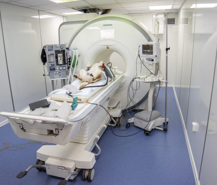
CT can offer the Veterinary profession superior diagnostic imaging capabilities in areas of the body that are difficult to image by other means.
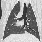
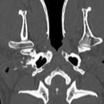
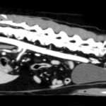
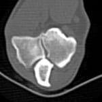
CT Clinical Gallery
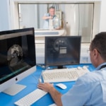
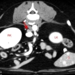
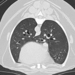
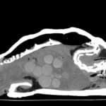
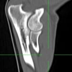
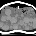
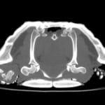
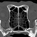
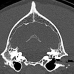
Images are acquired in one plane- axial, so the patient is scanned once, then using complex computer algorithms the data generated from this scan is used to create images in dorsal, sagittal or oblique planes or even 3D to allow for more precise pinpointing of lesions. This process is called either reformatting, reconstructing or re-slicing and the images produced are called multi-planar reformats or reconstructions or MPRs. The ability to view in multiple panes aids in prognostication and surgical planning.
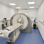
Adding IV contrast can also help further in identifying pathology. MPRs can be created on the CT workstation itself or on a desktop computer using third party software programs. CT is generally a more flexible technique than MRI- allowing imaging of head, thorax, abdomen and the entire skeleton, so is suitable for trauma, oncology, medical, orthopaedic and specific neurological cases. Patient sizes that can be imaged by CT range from the smallest exotic pet to full size equines if large bore CT is available. The CT gallery demonstrates the quality of image achievable using refurbished 16 or 64 slice CT systems. All images were acquired on systems installed in veterinary practices by Probo Veterinary.